
Case 145
- Wangpan Shi
- Jul 7
- 1 min read

A 54-year-old female with multiple bone lesions.


What's the molecular findings of this lesion?
A: FUS-CREB fusion
B: EWSR1-FLI1 fusion
C: FOS rearrangement
D: WWTR1-CAMTA1 fusion
E: EWSR1-SMAD3 fusion
Answer
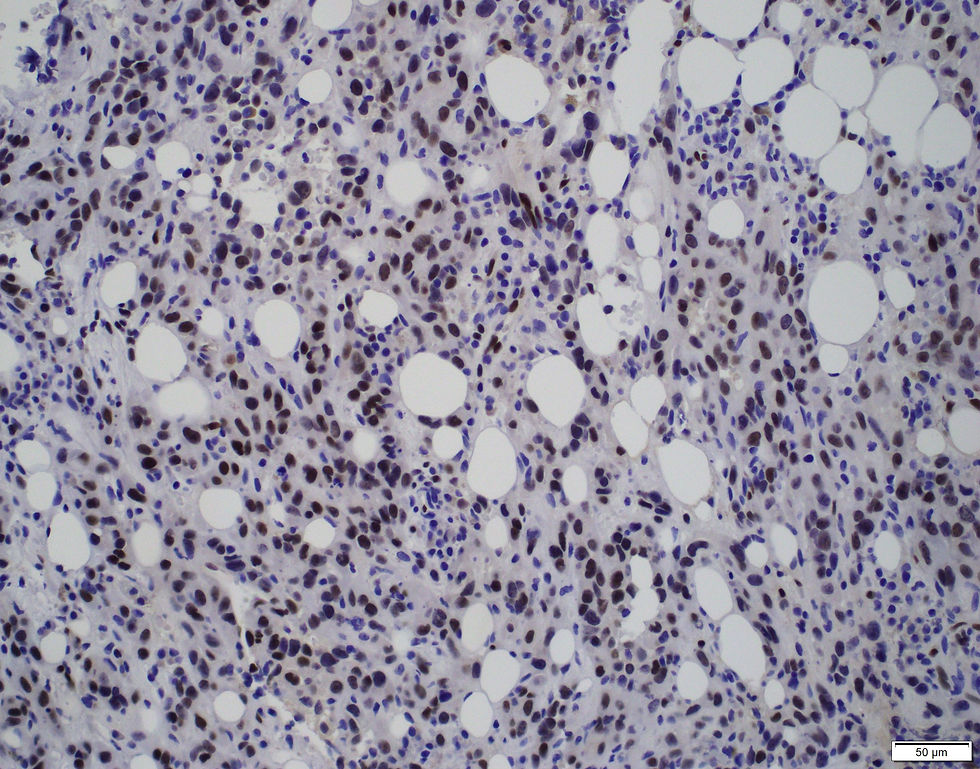
The correct answer is D. The immunostains are ERG and CD31. This is a case of epithelioid hemangioendothelioma in the bone.
Angiocentric tumours are characterized by their expansion of vessel walls, obliteration of the vascular lumen, and centrifugal infiltration into surrounding tissues, often provoking a sclerotic response and sometimes invading skeletal muscle or fat. Epithelioid haemangioendothelioma (EHE), a distinct vascular tumour, typically displays cords and nests of epithelioid cells embedded in a myxohyaline stroma. These tumour cells exhibit moderate eosinophilic cytoplasm, round nuclei with inconspicuous nucleoli, and may contain intracytoplasmic vacuoles occasionally housing erythrocytes.
Histologically, EHE usually presents minimal atypia and a low mitotic rate, although a small subset (less than 10%) shows atypical features such as nuclear pleomorphism, increased mitotic activity, necrosis, and sheet-like growth, potentially resembling epithelioid angiosarcoma.
Variant forms such as YAP1-TFE3 fusion-positive EHE exhibit brighter eosinophilic cytoplasm, solid growth, and frequent formation of vascular spaces—features generally absent in classic EHE.
Immunohistochemically, EHE expresses endothelial markers including CD34, CD31, podoplanin (D2-40), FLI1, and ERG, though staining intensity may vary. Approximately 40% of cases express epithelial markers like CK7, CK8, CK18, and pan-keratin, while EMA expression is rare. SMA is positive in about half of the cases.
Molecularly, EHE with WWTR1-CAMTA1 fusion shows strong nuclear CAMTA1 expression, whereas those with YAP1-TFE3 fusion demonstrate nuclear TFE3 expression, though TFE3 lacks specificity, as it can also appear in WWTR1-CAMTA1 fusion cases.
Case credit: UCSD Pathology
Author: Wangpan Jackson Shi, MD
Comments