
Case 47
- Wangpan Shi
- Feb 13
- 2 min read

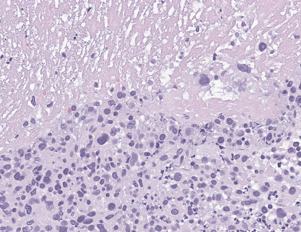
A 70-year-old women who presented with abnormal uterine bleeding and she underwent an endometrial biopsy. The tumor is strong and diffusely positive for p16.

What's your diagnosis of big category?
A: High grade serous carcinoma
B: High grade endometroid carcinoma
C: Sarcoma
D: Endometrial stromal sarcoma
E: Leiomyosarcoma
F: Poorly differentiated endocervical adenocarcinoma
G: Diffuse large B cell lymphoma
Answer
The correct answer is E, leiomyosarcoma and the diffusely p16 staining is not specific, p16 is frequently positive in high grade LMS. On the contrary, p16 loss is associated with CDKN2A deletion that can be associated with poor prognosis. The case showed tumor-type of necrosis and a high grade round, oval, spindle cell with cytoplasm. The leiomysarcoma was diagnosed based on diffuse positivity of desmin and negative for PAX8, keratin, wild type p53.
In low grade LMS, several mutations have been identified but none of them are diagnostic. Base on one paper of 77 tumors of leiomyoma, STUMP, leiomyosarcoma to study the recurrence:

Subtype | Histological Features | Mitotic Activity | Necrosis | Immunohistochemistry (IHC) |
Spindle Cell (Conventional) LMS | - Fusiform cells with eosinophilic cytoplasm in interlacing fascicles. - Nuclear pleomorphism varies, some cases appear uniform. - Multinucleated and osteoclast-like cells may be present. | High (≥4 mitoses/mm²) (≥10 mitoses/10 HPF). | Present in ~1/3 of cases - Abrupt transition from viable to non-viable cells. | Positive: SMA, Desmin, H-Caldesmon - ER/PR expression in spindle cell type - p16 and p53 overexpression common |
Epithelioid LMS | - Round/polygonal cells with eosinophilic or clear cytoplasm. - Nests, cords, nodules, or diffuse patterns. - Rhabdoid or signet-ring morphology may be seen. - Pseudoglandular spaces and hyalinization may occur. | ≥1.6 mitoses/mm² (≥4 mitoses/10 HPF). | May be present | Positive: CD10, EMA, Cytokeratin - Frequent positivity in epithelioid LMS |
Myxoid LMS | - Paucicellular with abundant myxoid stroma. - Fascicular or nodular growth may be seen. - Requires extensive sampling to identify malignant areas. | >0.4 mitoses/mm² (>1 mitosis/10 HPF). | Present | Patchy or weak SMA, Desmin, H-Caldesmon - May show p53 overexpression |
Other Features (Across All Subtypes) | - Mixed morphologies are common. - Infiltrative margins and vascular invasion in ~20% of cases. | Variable | Frequent in aggressive cases | Variable ER/PR expression in spindle LMS - p16 overexpression common in high-grade LMS |
Croce S, Ducoulombier A, Ribeiro A, Lesluyes T, Noel JC, Amant F, Guillou L, Stoeckle E, Devouassoux-Shisheboran M, Penel N, Floquet A, Arnould L, Guyon F, Mishellany F, Chakiba C, Cuppens T, Zikan M, Leroux A, Frouin E, Farre I, Genestie C, Valo I, MacGrogan G, Chibon F. Genome profiling is an efficient tool to avoid the STUMP classification of uterine smooth muscle lesions: a comprehensive array-genomic hybridization analysis of 77 tumors. Mod Pathol. 2018 May;31(5):816-828. doi: 10.1038/modpathol.2017.185. Epub 2018 Jan 12. PMID: 29327710.
Case credit: UCSD Pathology
Author: Wangpan Jackson Shi, MD
Comments