
Case 48
- Wangpan Shi
- Feb 14
- 2 min read

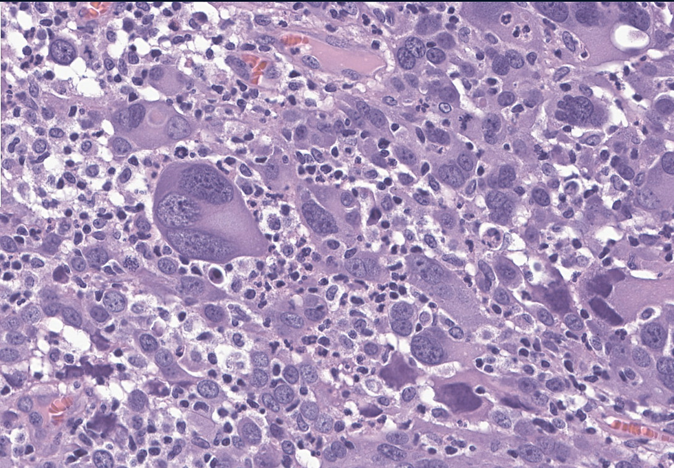
A 45-year-old female with an endometrial mass.


What's the diagnosis?
A: High grade endometrioid carcinoma, FIGO3
B: Mixed endometrioid and high grade serous carcinoma
C: High grade serous carcinoma
D: Giant cell carcinoma of endometrium
E: Mixed high grade endometrial endometrioid and giant cell carcinoma
Answer
The correct answer is E. This is a very rare case that only described in case reports and not recognized in WHO. Immunohistochemical studies performed show tumor cells to be positive for PAX8, EMA, pankeratin and E-cadherin (patchy) while to be negative for HER2 (score 0, no staining), Napsin-A, ER, PR, GATA3, and CD68. p16 is strong diffusely positive in tumor cells. PTEN and PAX2 expressions are lost and beta catenin does not show nuclear expression. p53 staining pattern is equivocal but most areas appears to be wild type. p63 shows focal staining. Overall, the pathologic findings are consistent with mixed endometrial carcinoma with high grade endometrioid carcinoma and giant cell carcinoma components
Category | Findings | Key Insights |
Patient Demographics | Ages: 55 to 76 years | All cases were confined to the uterus (FIGO IA or IB). |
Histological Features | - Dyshesive bizarre giant cells with atypical mitoses. - Minor components: endometrioid, serous, spindled/myxoid, and undifferentiated areas. - High mitotic index (43–211 mitoses/10 HPF). | Highly pleomorphic tumor cells with extensive mitotic activity and mesenchymal differentiation in some cases. |
Immunohistochemistry (IHC) | - Positive: Cytokeratin AE1/AE3, EMA (focal/multifocal), p16 (diffuse), hormone receptors (focal/multifocal), vimentin (2/3 cases). - Negative: E-cadherin (loss), CD68, α-FP, β-HCG, muscle markers (desmin, SMA, calponin), CD10, ERG. | - Partial loss of epithelial markers. - p53-abnormal staining in 1 case. - Mismatch repair-proficient and microsatellite-stable in all cases. - No POLE mutations detected. |
Molecular Findings | - TCGA classification: - 2 cases = No Specific Molecular Profile (NSMP). - 1 case = p53-abnormal. | - Unlike undifferentiated/dedifferentiated carcinoma (UDEC/DDEC) or carcinosarcoma (ECS), EGCC is mostly NSMP. |
Differential Diagnosis | - Lack of histiocytic (CD68-) and muscle markers (-SMA, -Desmin, -H-Caldesmon) excludes other mimics. - Partial loss of epithelial markers but focal cytokeratin positivity supports carcinoma. - No POLE mutations differentiate from UDEC/DDEC. | - Resembles UDEC/DDEC and carcinosarcoma but lacks consistent p53 mutations and MMR deficiency. |
Treatment & Outcome | - All patients received carboplatin + paclitaxel chemotherapy + radiotherapy. - All were alive with no evidence of disease at 12, 8, and 3 months follow-up. | - Aggressive features but uncertain long-term prognosis due to limited cases. |
Clinical Significance | - EGCC should be considered a variant of high-grade endometrial carcinoma rather than a distinct entity. - May require management similar to undifferentiated or carcinosarcoma. | - Further studies needed to determine prognosis and molecular classification. |
References: Arciuolo, D., Travaglino, A., Raffone, A. et al. Endometrial giant cell carcinoma: new insights from a morphological, immunohistochemical, and molecular analysis of three cases. Virchows Arch 481, 321–326 (2022). https://doi.org/10.1007/s00428-022-03310-x
Case credit: UCSD Pathology
Author: Wangpan Jackson Shi, MD
Comments